The Medical Analysis Department at the Faculty of Applied Science, Tishk International University, successfully completed a comprehensive six-session hands-on training course on molecular biology techniques, designed to integrate theoretical knowledge with laboratory practice.
The program commenced with the first session that was held on the 25th of January 2026, which was fully theoretical. This opening lecture introduced students to the course outline, laboratory safety measures, and the fundamental principles of molecular biology that would guide the subsequent practical work.
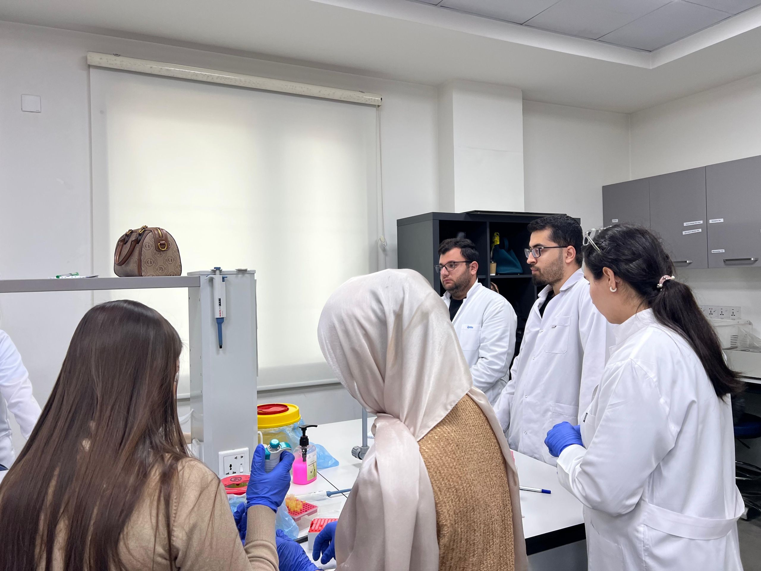
During the second session that was held on the 26th of January 2026, students conducted hands-on genomic DNA extraction from human samples under the supervision of Dr. Sami Mamand, the head of the medical analysis department; Dr. Galawezh Obaid; and Dr. Jaafaru Sani, academic staff at TIU. Trainees performed the full extraction procedure, including sample processing, cell lysis, DNA isolation, and purification. Emphasis was placed on sterility and correct laboratory handling techniques.
The third session that was held on 1st of February 2026, returned to a theoretical format, providing an explanation of the Polymerase Chain Reaction (PCR). Trainees were introduced to the mechanism of PCR, its essential components, and the different types of PCR techniques used in molecular diagnostics and research. The session detailed how thermal cycling enables DNA amplification.
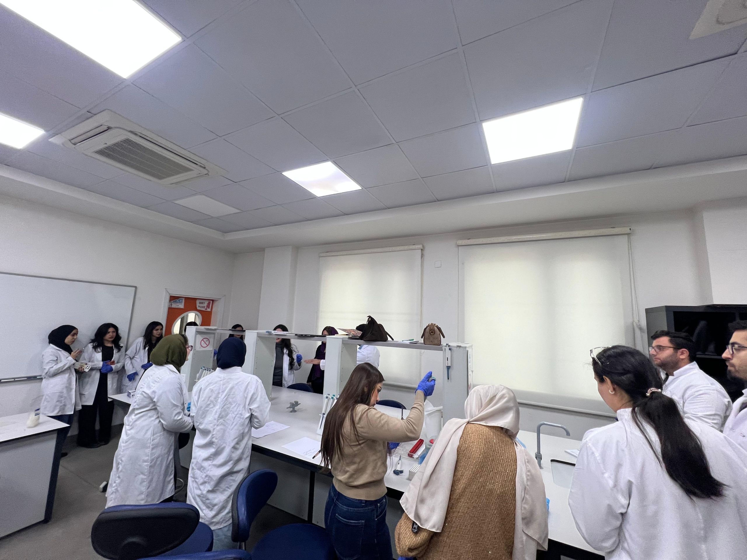
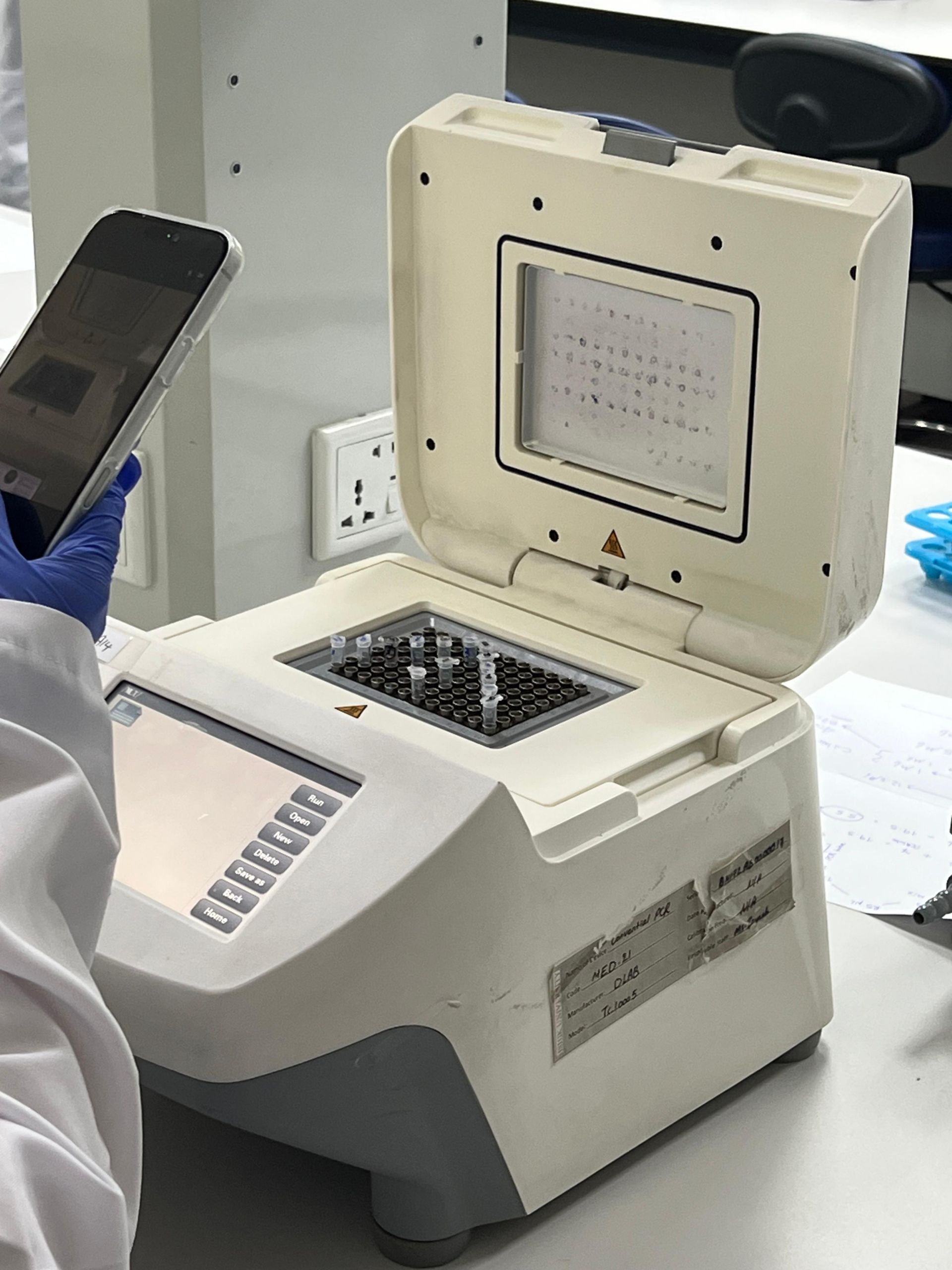
In the fourth session that was held on 2nd of February 2026, trainees performed PCR amplification. They prepared master mixes, placed the reagents into PCR tubes, and operated the thermocycler to amplify their previously extracted DNA samples.
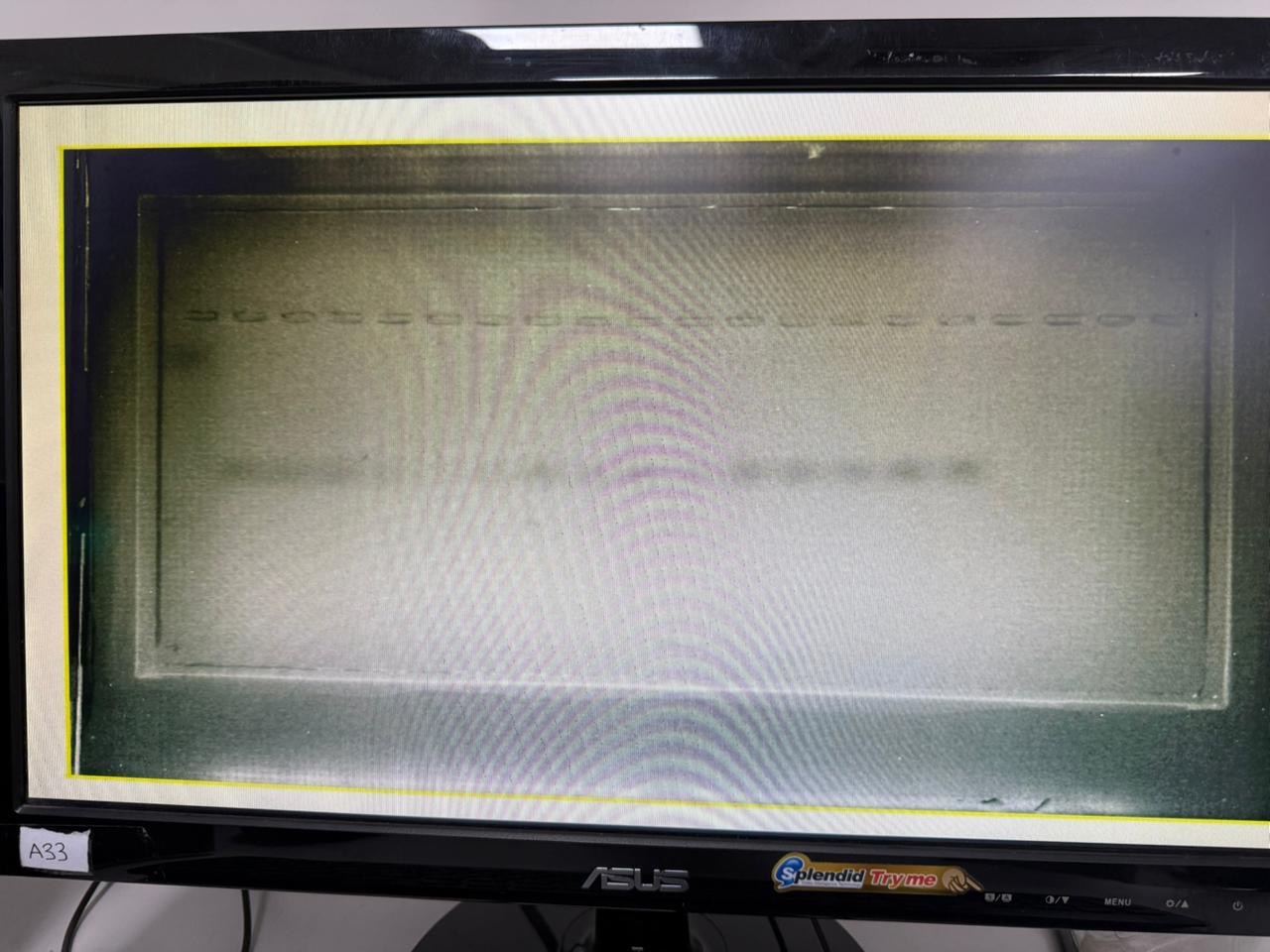
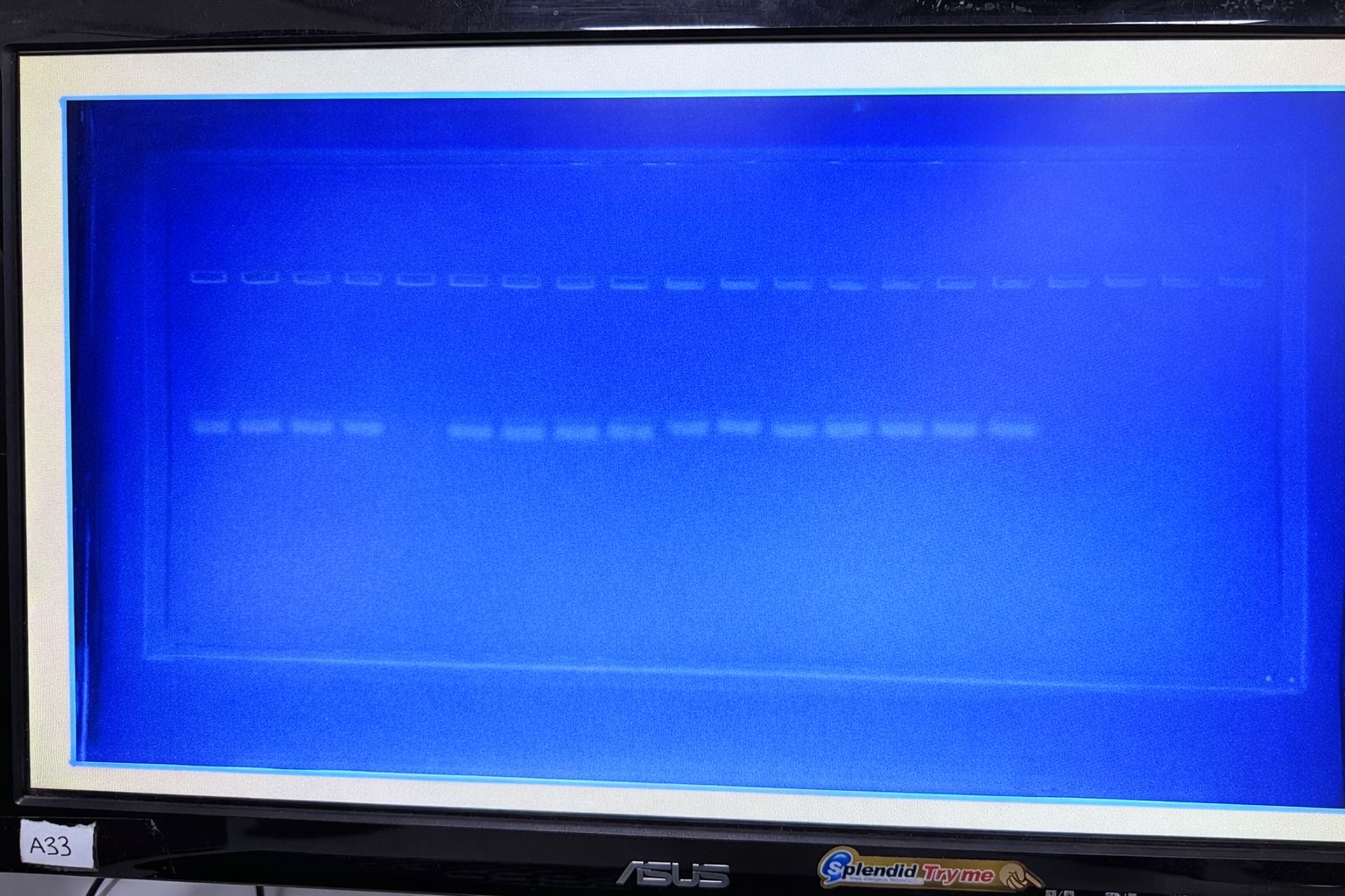
The fifth session that was held on 8th of February 2026, focused on agarose gel electrophoresis from a theoretical and preparatory perspective. Students learned how to prepare gels, the differences between gel concentrations and their impact on DNA fragment separation, the purpose of electrophoresis, and the types of DNA ladders used for molecular weight estimation. Detailed explanations were also provided regarding gel visualization techniques.
Finally, in the sixth session that was held on 9th of February 2026, students carried out gel electrophoresis using their own PCR products. They loaded samples into prepared agarose gels, performed electrophoretic separation, and visualized the amplified DNA fragments using a UV transilluminator. The session concluded with gel documentation, enabling students to record and analyze their experimental results.
Through this structured six-session program, participants completed the full molecular workflow, from theoretical lectures to DNA extraction, PCR amplification, gel electrophoresis, visualization, and documentation, reflecting the department’s ongoing commitment to strengthening practical and academic excellence.